
What is Chromosomes?
- Chromosome is a microscopic thread like structure.
- It is a part of cell that carry hereditary information in the form of genes.
- Chromosome are the rod shape, dark stained bodies which is seen only at metaphase stage of mitosis.
- Strausberger in 1875 was first to describe chromosome and word chromosome was given by Waldeyer in 1888.
Structure of Chromosome
A chromosome is a long, continuous strand of DNA containing numerous genes, regulatory elements, and other nucleotide sequences. In eukaryotes, DNA is coiled around histone proteins, forming a compact structure, whereas in prokaryotes, chromosomes are typically circular and lack histones.
Structure of a Eukaryotic Chromosome
A. Chromatin
- DNA + histone protein complex.
- Exists in two forms:
- Euchromatin – lightly packed, transcriptionally active.
- Heterochromatin – tightly packed, transcriptionally inactive.
B. Nucleosome
- Fundamental unit of chromatin.
- DNA (~147 base pairs) wrapped around a histone octamer (2 each of H2A, H2B, H3, H4).
C. Solenoid/30 nm fiber
- Nucleosomes coil further into a thicker fiber, stabilizing the structure.
D. Scaffold and Supercoiling
- Higher-order folding around a protein scaffold leads to the metaphase chromosome.
Structure of Prokaryotic Chromosome
- Typically single circular DNA molecule
- Found in the nucleoid region
- May contain plasmids – small, circular, extra-chromosomal DNA
- Lacks histones (except some archaea)
Chromosome number and size
- Each species has a somatic and gametic chromosome number.
- Somatic chromosome number is the number of chromosomes present in somatic cells and it is shown by 2n.
- Gametic chromosomes number is the number of chromosomes present in gametic cells of an organism. It is shown by n and is precisely one half of the somatic number.
- Researchers define homologous chromosomes as the two copies of a chromosome, which are identical in morphology, gene order, and gene content.
- The size of chromosomes, depends on the different stages of the cell division.
- During interphase, chromosomes are not visible under light microscope because at this stage they are longest and thinnest, but in other phase chromosomes are visible due to progressive decrease in length and increase in thickness.
Morphology
- Morphology of chromosomes will be change at the stage of cell division, and metaphase chromosomes are foremost suitable for studies.
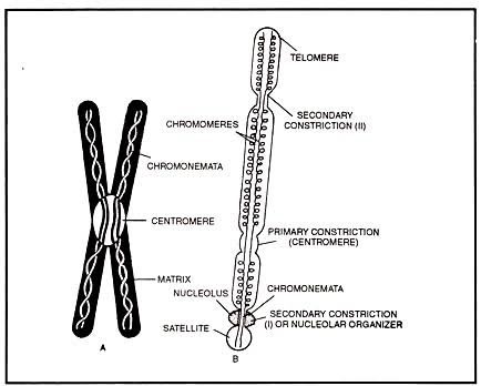
The following structural features are seen under the microscope, in metaphase chromosome:
-
- Chromatid
- Centromere
- Telomere
- Secondary constriction and satellite chromosomes.
- Chromomere (seen during prophage).
Chromatid
- Each metaphase chromosome divides into two identical parts (longitudinally), and we refer to each part as a chromatid.
- Chromatid is that the structural and functional unit of chromosomes and its not further subdivided into smaller units.
- Sister chromatids: The two chromatids building up a chromosome develop from the replication of one chromatid.
- The chromatids of homologous chromosomes are called non-sister chromatids.
Centromere
- The two chromatids of a chromosome seem to join or fuse in the region of the centromere.
- Under the microscope, scientists generally observe the centromere as a constriction (a narrowed region) in the chromosome, hence they know it as Primary Constriction.
- The centromere splits each chromosome into two transverse parts: these parts are known as Arms.
- In most cases, one arm of the chromosomes is longer than the other, hence they’re termed as Long arm and short arm, respectively.
- Long is represented by q while short arm usually denoted by p.
On the basis of position of their centromere, chromosome is of 4 types:
-
- Metacentric chromosomes
- Submetacentric chromosome
- Acrocentric chromosome
- Telocentric chromosome
Metacentric chromosome
- Centromere is found at the center of chromosome.
- Centromere is median.
- Two arms of such chromosomes are equal and ARM ratio is 1.
- Such chromosomes appear V-shaped, during anaphase.
Submetacentric chromosome
- Centromere is submedian and located on one side of the central point.
- Chromosomes appear either like ‘V’ or ‘J’ shaped, during anaphase.
Acrocentric chromosome
- They are subterminal.
- Centromere are located near one end of the chromosome.
- Chromosomes may appear either like ‘J’ or rod-shaped, during anaphase.
Telocentric chromosome
- Centromere located at one end of chromosomes.
- Such chromosomes are called terminal.
- They always appear as rod- Shaped, during anaphase.
Monocentric chromosome: chromosomes in most species features a single centromere.
Polycentric chromosome: Each chromosomes has more than one centromere.
Telomere

- The two ends of a chromosomes are known as telomere.
Secondary constriction or satellite chromosome
- In some chromosomes, is second constriction, is present additionally to the primary constriction, this extra constriction called, secondary constriction.
- Scientists know the region between the secondary constriction and the nearest telomere as satellite.
- They call chromosomes having secondary construction satellite chromosomes or sat-chromosome.
Chromomere
In some species E.g. maize, amphibia etc. chromosomes during the first prophase of meiosis, more particularly during pachytene, show small bead like structure called chromomere.
Chromatin
- It is material of which chromosomes are composed.
- Chromatin are classified into two groups, on the premise of stainability with basic dye:
-
- Heterochromatin
- Euchromatin
Heterochromatin
- Take deep stain during prophase and interphase.
- Lightly stained, during metaphase.
Euchromatin
- Takes up little stain during interphase, stains lightly during prophase.
- Stained deeply, during metaphase.
Heterochromatin is divided into two groups:
- Constitutive
- Facultative
Constitutive heterochromatin
- Remains permanently within the heterochromatic state.
- It doesn’t revert back to the Euchromatin State. E.g. – centromeric region.
Facultative heterochromatin
- Euchromatin undergoes hetero chromatinization (conversion into heterochromatin).
Chemical composition
- Chromatin is consisting of DNA, RNA and Protein.
- Chromatin isolated from interphase nuclei contain:
-
- 30-40% – DNA
- 50-65% – Protein
- 0.5-10% – RNA
- Metaphase chromosomes contain:
-
- 15-20% – DNA
- 10-15% – RNA
- 65-70% – Protein
Organization of chromatin fiber (nucleosome solenoid model)
- Both metaphase chromosomes and interphase nuclei display fibers of 200-300 A°; researchers call these fibers chromatin fibers.
- Chromatin fibers are the basic and essential unit of chromosome structure.
- The nucleosome solenoid model of chromatin fiber is universally accepted.
- Kornberg and Thomas developed this model in 1974.
- According to this model, chromatin is consisting of a repeating unit called Nucleosome.
- One complete nucleosome, a whole disc of 11nm diameter and 6nm height.
- Nucleosome contain an average of ~200bp DNA which usually ranges between 180 and 200bp.
Nucleosomes consists of:
-
- Nucleosome core
- Linker DNA
- H1 Histone
- Other chromosomal protein
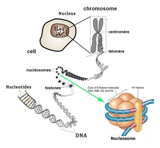
Nucleosome core
- It consists of histone octamer, composed of two molecules each of histones H2a, H2b, H3 and H4.
- In addition, this histone octamer winds a 146 bp long DNA molecule round itself in 1-3/4 turns.
- This segment of DNA in chromatin fibers is nucleus resistant.

Linker DNA
- The size of linker DNA vary from 8 bp to 114 bp depending on the species.
- This DNA forms the string a part of the heads on a string structure seen on isolation of chromatin fiber, and is nuclease susceptible.
H1 histone
- Each nucleosome consists of an average one molecule of H1 histone.
- The complete removal of H1 histone leaves the structure of the nucleosome core unaffected, showing that H1 resides outside the nucleosome core.
Other chromosomal proteins
- Both linker DNA nucleosome are related with other chromosomal protein.
- In native chromatin, the beads are about 110A° in diameter is 60A° high and ellipsoidal in shape.
- The nucleosome stacked together without any detectable linker DNA between, this produces the 100A° thick chromatin fiber called nucleosome fiber.
- The Nucleosome fiber is also called 10 nm fiber.
- The nucleosome fiber may then supercoil to give rise to the 300A° chromatin fiber seen in electron micrographs of metaphase chromosome.
- Researchers call the supercoiled nucleosome fiber 30nm fiber, which most likely exhibits solenoid organisation of nucleosome.
Reference and Sources
- 1% – https://onlinesciencenotes.com/chromosomes/
- 1% – http://www.authorstream.com/Presentation/suvarna118329-2237733-special-type-chro
mosome/ - 1% – https://www.yourarticlelibrary.com/biology/physical-basis-of-life-chromosome-appearance-morphology/41736
- 1% – https://www.thoughtco.com/diploid-cell-373464
- 1% – https://www.biologydiscussion.com/chromosomes/chromosomes-introduction-number-and-types-cell-biology/26817
- 3% – https://www.yourarticlelibrary.com/biology/chromosomes-structure-functions-and-other-details-about-chromosomes/22946
- 1% – https://www.slideshare.net/AashishPatel14/chromosome-and-its-structure
- 1% – https://www.biologydiscussion.com/chromosomes/chromosomes-in-the-nucleoplasm/36998
- 1% – https://www.slideshare.net/tvr329/chromosome-structure-function
Also Read:
- Vector: properties, types and characteristics
- What is Gene Expression?
- DNA replication in prokaryotes
- Mutations: Introduction, Types, Causes and Repair Mechanisms
- Microorganisms in Benthic Marine Environments
- Probiotics: Introduction, Development and Uses in Agriculture
- Major histocompatibility complex (MHC): introduction, types and differences
- DNA Replication in eukaryotes: Initiation, Elongation and termination
- Hansen’s Disease: Symptoms, Causative Agent, Pathogenesis and Treatment
- CRISPR-Cas9 Gene editing tool: Introduction, Principles, Uses & Applications
- Microbial Identification and Strain Typing Using Molecular Techniques
- Reverse Transcription Polymerase Chain Reaction (RT-PCR)
- Microbiology Disciplines: Bacteria, Viruses, Fungi, Archaea and Protists
- Fungi: Distribution, Morphology, Reproduction, Classification

7 thoughts on “Chromosomes: Structure, Morphology, Composition and Organization”
I liked these concepts very much, it’s easily to understand and easy to learn
thank you ankita keep supporting us
How to download them as pdf
Thank you so much mam ,
It’s remembered me, when i was high school. I liked it.
Thank you so much, keep supporting us
it’s too much helpful 4 students
i really like it the way this website explain each and every point.