
Introduction
- Staining methods are used to elevate the visibility and highlight specific morphological structures of the microorganisms. It is also used to preserve them.
- Before staining, the specimen is fixed by the method of fixation.
- Fixation is a method in which the complete structure of the cells or microorganisms are fixed in the positions and are preserved.
- Inactivation of enzymes, which cause cell morphological disruption.
- Cell structure gets rigid in form, which helps it to remain stationary during staining & observation.
- Usually, microorganisms are killed & attached strongly to the slide during the fixation process.
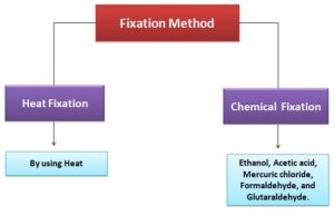
| Fixation Methods | Remarks |
| Heat Fixation | Used to observe prokaryotic cells
Smear is prepared and passed through the flame, which is gently heated. Preserve the whole morphology of the cells, but not the internal structure within the cell |
| Chemical Fixation | Used to protect cell morphology as well as cell fine substructures of the more delicate microorganisms
Chemical fixative gets inside the cells & reacts with components present in the cells, such as proteins & lipids to immobile them. |
Common features of Staining Methods
There are various dyes used for staining, have the following features in common:
- Chromophore groups: It has a conjugated double bond, binds with cells by covalent, ionic, or hydrophobic bonding.
- It gives dye its color.
Different types of Dyes which are used for staining
- Usually, dyes directly stain the cell, but few dyes stained the background, not the cells such as Indian Ink & Nigrosin, the method is known as negative staining, in which the cells appear to be bright against the dark background.
- Commonly used dyes bind cells by ionic bonding, and these ionizable dyes are divided into two classes on the basis of charge:
- Basic dyes
- Acidic dyes
| Dyes | Examples | Remarks |
| Basic Dyes |
Basic Fuchsin Methylene Blue Crystal Violet Malachite Green Safranin |
These dyes are positively charged (a form of pentavalent nitrogen), commercially available as chloride salts.
It forms a bond with negatively charged molecules for instance nucleic acids, proteins & even the cell surface of single-celled microorganisms. |
| Acidic Dyes | Rose Bengal
Eosin Acid Fuchsin |
These dyes are negative charge, it binds to cell structures which are positive in charge. |
- NOTE: Staining ionizable dyes effectiveness varies with change in pH which is associated with alteration of charge on cell molecules. Thus acidic conditions favor the work of acidic dye stain whereas alkaline pH works well with basic dyes.
- Based on the solubility characteristics of the dyes, they form covalent bonds with cellular components.
- In Feulgen Procedure, Schiff’s reagent covalently bonds with deoxyribose sugars and stain the DNA.
- Lipid soluble Sudan III (Sudan Black), stain lipids.
Different Types of Staining Methods
There are several staining methods which are all mentioned below:
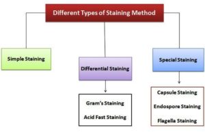
Simple Staining
- Simple staining is used to stain prokaryotic cells for e.g., microorganisms, used to determine the shape, size, and arrangements.
- In this staining method, only a single dye is needed.
- It’s simple with the procedure which includes covering fixed smears with stain for a minute, and excess stain is washed off with water and blotted dry.
- In this case, basic dyes are used which are methylene blue, crystal violet, and carbolfuchsin.

Differential Staining
Gram Staining
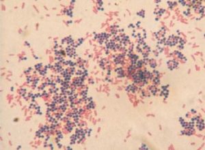
- Very commonly used differential staining is Gram staining, used to differentiate gram-positive & gram-negative bacterias.
- Developed by Christain Gram in 1884
- Procedure:
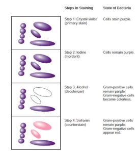
- The gram-positive bacteria have a cell wall composed of thick layers of peptidoglycan, during the decolorizing process in step 3, in order to remove the stain, alcohol shrinks & dehydrate the cell which leads to the closing of pores on the cell wall, through which primary stain is not able to escape and they remain purple.
Acid Fast Staining
- Acid Fast Staining is also known as the Ziehl Neelsen method, commonly used to identifies Mycobacteria spp i.e., M. tuberculosis & M. leprae. and helps to differentiate acid-fast bacteria & non-acid fast bacteria.
- The cell wall of these bacteria consists of large content of lipid, especially mycolic acid ( branched-chain of hydroxy lipid), which prevents dye-binding.
- In this staining procedure, primary stain Basic Fushcin gets into the cells with the help of prior heat treatment.
- When primary stain penetrates the cell of acid-fast bacteria, for e.g., Mycobacterium, acid alcohol won’t be able to decolorize them.
- Therefore acid-fast bacteria appear red, whereas non- acid-fast bacteria appear blue because of counterstain methylene blue.


Special Staining
- This staining method is used to identifies or study special components of the microorganisms.
| Special Staining | Procedure | Remarks |
| Capsule Staining |
|
|
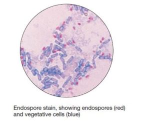
| Endospore staining |
|
|
| Flagella staining |
|
|

References and Sources
- https://imicrobiology.blogspot.com/2020/12/microscopy-specimen-preparation-bright-field-dark-fieldphase-
contrast-fluorescent-electron-microscope.html - https://www.researchgate.net/publication/7666663_Safe_Specimen_Preparation_for_Electron_Microscopy_of_
Pathogenic_Fungi_by_Freeze-substitution_after_Glutaraldehyde_Fixation - https://www.easynotecards.com/print_cards/79794
- https://www.foodcolorworld.com/basic-dyes.html
- https://nios.ac.in/media/documents/dmlt/Microbiology/Lesson-02.pdf
- https://www.studymode.com/essays/Gram-Staining-Lab-Report-85886659.html
Also read
- Gram staining
- Nitrogen Cycle
- Measurements of microbial growth
- Plasma Membrane- Structure, Properties, Lipid Rafts and Glycolipids
- Microbial Identification and Strain Typing Using Molecular Techniques
- Endospore staining and capsule staining of the bacteria
- Electrophoresis: Overview, Principles and Types

1 thought on “Different Staining Methods used in Microbiology”
Very helpful ! Easy to understand. Thank u .