
Microscopy: Overview, Principles and Its Types
Principles of Microscopy
- Biochemical analysis is generally go along with microscopic investigation of cell, tissue or organelle preparations. Such type of investigation are used in various applications, for e.g. for the biochemical characterization or to examine the integrity of samples during the experiment.
- Microscopy is a technique use for making very tiny things to visible to the naked eyes and the instrument used to make things visible to the unaided or naked eye is known as Microscope.
- The microscope is the instrument most frequently characteristics of microbiology laboratory.
- The magnification provides by the microscope enables us to see microorganisms and their structures otherwise invisible to the naked eye.
- There are two fundamental types of microscope:
- Light microscope
- Electron microscope
- Light microscope involves the use of series of glass lenses to focus light in order to create an image whereas, electron microscope uses electromagnetic lenses to focus beam of electrons.
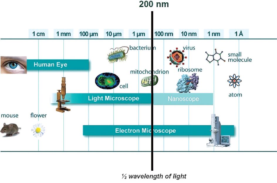
- Light microscopes have ability to magnify objects up to a maximum of approximately 1500 times whereas electron microscopes are able to magnify the objects maximum of approximately 200,000 times.
- Magnification is not the best measure of microscope, however. Rather, resolution, that is the ability to distinguish between two closely spaced objects in a specimen which is the more reliable estimate of a microscope’s utility.
- Resolution limit of standard light microscope is about 0.5 micrometers but in contrast lateral resolution limit of electron microscope is up to 1 nanometer (nm).
- Light microscopes are used to view both living and dead specimens and often in real color, but in electron microscopes only dead specimens are viewed and never in real color.
Light microscope
- It is a type of microscopy in which magnifications is obtained by a system of optical lenses using light waves.
- Light microscope abide of a single lens mounted in a metal frame is the simple form of microscope- a magnifying lens.
- Light microscope usually uses sun or ambient indoor light as a source of illuminations.
- Because of the travelling of light through the specimens, this instrument is also called as transmission light microscope.
- The light microscope forms a magnified image of a specimen which is based on the principles of absorption, transmission, diffraction and refraction of light waves.
- All modern light microscopes are usually made up of more than one glass in combinations in which the major components are the eyepiece lens or ocular lens, the objective lens, and the condenser lens, and instruments of such combinations are therefore called compound microscopes.
Types of light microscopes
- Bright field microscope: In this type of microscopy, the area observed or the microscopic field is appears brightly lighted whereas the microorganisms appears dark because they absorb some of the light.
- Dark filed microscope: In this, effects produced by the dark field techniques is that of a dark background against which objects are brightly illuminated.
- Phase contrast microscope: In principle, this technique is based on the fact that light passing through one material and into another material of a slightly different refractive index and/or thickness will undergo in change in phase.
- Fluorescence microscope: In this the specimens itself acts as a light source, the specimens used to study are either fluorescent materials or stained with fluorescent dye.
Electron microscope
- Electron microscope is used when the greatest resolution is required, and when the living state can be ignored.
- The image produced in an electron microscope reveals the ultra structure of cells.
- The source of light in an electron microscope is the electron gun or electron beam.
- A tungsten filament emits electrons, when a high voltage of between 40 000 and 100 000 volts (the accelerating voltage) is passed between the cathode and the anode.
Types of electron microscope
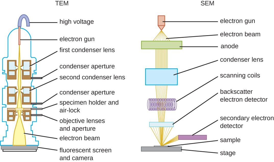
Transmission electron microscope (TEM): In the TEM, electrons that passes through the specimen are imaged.
Scanning electron microscope (SEM): In the SEM electrons that are reflected back from the specimen (secondary electrons) are collected, and the surfaces of specimens are imaged.
Also Read:
- Simple Microscope
- Types of microscopes
- Chromatography: Introduction, Principle, Classification and applications
- Antibody or Immunoglobulin
- Microbiology Disciplines: Bacteria, Viruses, Fungi, Archaea and Protists
- Fundamental Principle of Clinical Specimen Collection
- Second Golden Age of Microbiology
- Methods for Laboratory diagnosis of viral infections
Microscopy: Overview, Principles and Its Types
Reference and Sources:
- https://www.researchgate.net/publication/322208919_Microscopy
- https://idoc.pub/documents/textbook-of-practical-microbiology-pon28529kjn0
- https://www.cambridge.org/core/books/wilson-and-walkers-principles-and-techniques-of-biochemistry-andmolecular-biology/microscopy/A04B7CC4399463585DC7D6AF1A6523FC
- https://www.scribd.com/presentation/80195320/The-Microscope