
Introduction
Microscopes are essential tools for visualizing objects too small to be seen with the naked eye. They are classified based on the source of illumination (light or electron), the specimen type, and the image type produced.
Light microscope
- Light microscopy in which magnifications is obtained by a system of optical lenses using light waves.
- Microscope contains a single lens mounted in a metal frame which is the simple form of microscope- a magnifying lens.
- Light microscope always uses sun or ambient indoor light as a source of illuminations.
- Because of the traveling of light through the specimens, this instrument is also called as transmission light microscope.
- The light microscope obtains a magnified image of specimen which is based on the principles of absorption, transmission, diffraction and refraction of light waves.
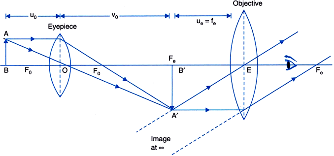
- All modern light microscopes are mainly made up of more than one glass in combinations in which the major components are the eyepiece lens or ocular lens, the objective lens, and the condenser lens, and instruments of such combinations are therefore called compound microscopes.
- Condenser lens focuses the light at the specimen from the light source.
- The one facing the object is called objective lens and the one close to the eye is called the eyepiece.
- It is objective lens that is responsible for producing magnified images.
- Objective lens is available in different magnifications such as 4x, 10x, 20x, 40x, 60x, 100x.
- Eyepiece have larger aperture and larger focal length than the objective lens.
- Objective lens and eyepiece both work in combination to magnify the objects.
- A compound microscope with single eyepiece is termed as monocular whereas, with to eyepiece is termed as binocular.
- The part of the microscope that holds all of the components firmly in position is called the stand.
- There are two types of compound microscope stand- an upright or an inverted microscope.
- In an upright microscope, light source is below the condenser lens and the objective lens is above the specimen stage, whereas in inverted microscopes, the condenser lens and the light source is above the specimen stage and objective lens beneath it.
Magnification
- The magnification or linear magnification is defined as the ratio of the image size to the object size.
- Both magnification and magnifying power is the different terms, magnifying power or angular magnification is the angle subtended by object and the image.
- Magnification of objective lens is called the linear magnification, while magnification of the eyepiece is called the angular magnification. So, the total magnification of the compound microscope is the is product of both the linear magnification of the objective lens and the angular magnification of the eyepiece.
Resolving power
- It may be defined as the ability of microscope to distinguish two objects that are close together.
- It is inversely proportional to the limit of resolution and the limit of resolution is defined as the minimum distance between the two points that allows their discrimination as two separate points.
Types of light microscope
- Bright field microscope
- Dark filed microscope
- Phase contrast microscope
- Fluorescence microscope
Bright field microscope
- It is most basic mode of the light microscope, which can be viewed with the minimum optical elements.
- In this type of microscopy, the area observed or the microscopic field is appears brightly lighted whereas the microorganisms appears dark because they absorb some of the light.
- In the bright field of microscopy specimen is viewed by transmitted light from the condenser lens.
- This microscopy, light is directed towards the condenser lens, through the specimen, through an objective lens, and to the eye through the ocular lens or eyepiece (a second magnifying lens).
- Bright field microscope is most commonly used to collected images of histological sections or pigmented tissue or tissue culture cells that have been stained with colorful dyes.
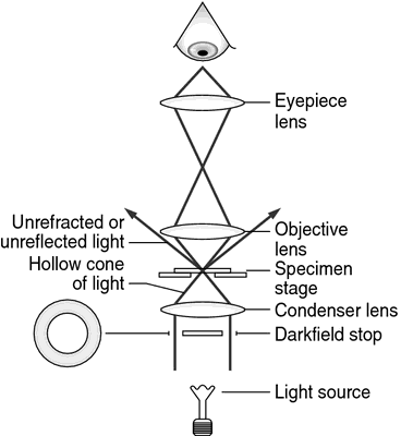
Dark field microscope

- In this, effects produced by the dark field techniques is that of a dark background against which objects are brightly illuminated.
- This technique is most often used for improving the contrast if unstained, transparent specimens.
- This is accomplished by placing an opaque disc underneath the condenser lens, that blocks the direct entering of light in the objective lens; light reflected by the specimens enters the objective and the specimen appears light against a black background.
- This technique has been used traditionally for viewing the outlines of objects in liquid media such as microorganisms, spermatozoa or cells growing in the tissue culture or for quick check of the status of a biochemical preparation.
Phase contrast microscope

- This technique is generally based on the fact that light passing from one material and into another material of a slightly different refractive index and/or thickness will undergo in change in phase.
- It is most often, used for testing cell organelle preparations for lysis and for viewing unstained cells growing in tissue culture
- This method of microscopy images differences in the refractive index of cellular structures. Light which passes from the thicker parts of the cell is held up relative to the light that passes from thinner parts of the cytoplasm.
Fluorescence microscope
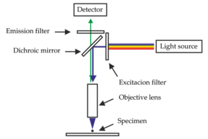
- In this the specimens itself acts as a light source, the specimens used to study are either fluorescent materials or stained with fluorescent dye.
- The fluorescence microscope is most often similar to the ordinary microscope except that the illuminating light is passed through two sets of filters.
- Excitation filter: It filters the light before reaches the specimen.
- Barrier or emission filter: It filters the light illuminated from the specimen.
- The excitation filter passes only the wavelength that excites the particular fluorescent dye, while the barrier blocks out this light and passes only those wavelengths emitted when the dye fluoresces.
Electron microscope
- Electron microscope is used when the maximum resolution is required, and when the living state can be ignored.
- The image produced in an electron microscope reveals the ultra structure of cells.
- The source of light in an electron microscope is the electron gun or electron beam.
- A tungsten filament emits electrons, when a high voltage of between 40 000 and 100 000 volts (the accelerating voltage) is passed between the cathode and the anode.
Types of electron microscope:
- Transmission electron microscope (TEM)
- Scanning electron microscope (SEM)
Transmission electron microscope (TEM)
- In this, a condenser lens focuses the electron beam onto the specimen and electrons are transmitted through the specimen.
- A heated tungsten filament in the electron gun which generates an electron beam that is then focused on the specimen by the condenser.
Scanning electron microscope (SEM)

- In the SEM, electrons which are reflected back from the specimen (secondary electrons) are collected, and the surfaces of specimens are then imaged.
- To produce an image, the Scanning electron microscope (SEM) scans a narrow, tapered beam of electrons back and forth over the specimen.
- When this beam strikes a particular area of the specimen, surface atoms discharge a tiny shower of electrons called secondary electrons, and these are trapped by a special detector.
- Secondary electrons then entering in the detector strike a scintillator which causing it to emit light flashes that a photomultiplier converts to an electrical current and amplifies.
- Then, the signal is sent to a cathode-ray tube and produces an image like a television picture, that can be viewed or photographed. The number of secondary electrons that reaches the detector depends on the nature of the specimen’s surface.
- When the beam of electrons strikes a raised area of specimen, a large number of secondary electrons enter into the detector; in contrast, some of the electrons escape a depression in the surface and reach the detector. Thus, the raised areas of specimen are appear lighter on the screen and depressions are darker.
- A realistic 3D image of the microorganism’s surface or specimen results. The actual in situ location of microorganisms in ecological niches such as the lining of the gut and the human skin and can be examined.
Also Read:
- Antibody or Immunoglobulin
- Hypersensitivity Reactions
- Gram staining
- Bacteria: Shape, Size, Structure and other Membrane
References and Sources:
- https://www.researchgate.net/publication/322208919_Microscopy
- https://www.slideshare.net/sanchuyadav/unit-2-microscopy
- http://drrajivdesaimd.com/2015/03/26/refractive-error/comment-page-9/
- https://readthebiology.blogspot.com/

4 thoughts on “Types of microscopes”
Good info..Better to add images of inverted and upright compound microscope..Thank u
Thanks for your appreciation
The explaination is just so good keep on doing the good job
Thanks for the application.