
Plasma Membrane- Structure, Properties, Lipid Rafts and Glycolipids
Introduction
- All cells including both prokaryotic and eukaryotic, are surrounded by a plasma membrane, which separates its internal contents from the extracellular environment, and defines the boundary of the cell.
- The plasma membrane is considered as the fundamental structure of cellular evolution, as it was thought first cell i.e., self-replicating RNA was arisen enclosed in the membrane of phospholipids (a constituent of plasma membrane).
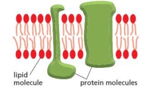
- All biological membranes have a common general structure, composed of a very thin film of lipid (fatty acid) and protein molecules (membrane protein), and held together by non-covalent interactions.
- Cell membranes are dynamic, fluid-structure and most of their molecules move in the plane of the membrane.
- The arrangement of lipid molecules is shown as a continuous double bilayer about 5nm thick. It serves as an impermeable membranes barrier to the passage of most water-soluble or polar molecules.
- But it is selectively permeable to organic molecules and ions, their movement across the membranes is mediated by membrane proteins (embedded in lipid bilayer), which act as a sensor, for extracellular signals.
- Hence, plasma membranes play a dual role, act as barriers and isolate the cytoplasm and mediate the cell interactions with its environment with the help of membranes proteins.
Chemical Constituents of Plasma Membranes
- All plasma membranes are composed of lipids and proteins, the ratio of protein to lipid varies enormously depends on different cell type. For e.g., human RBC contains 43% and 49% protein by weight, in contrast, mouse liver cells contain 54% and 46% protein by weight.
- The most abundant membrane lipids are the phospholipids composed of phosphoglycerides (have 3- C glycerol backbone), there are different phosphoglycerides formed by different cells, such as phosphatidylethanolamine, phosphatidylserine, and phosphatidylcholine.
- Another important phospholipid is sphingomyelin formed by sphingosine (long acyl chain with an amino group and two hydroxyl groups at the end of the molecule), rather than glycerol backbone.
- In eukaryotic plasma membranes, cholesterol (sterol) is found in large amounts.
- Plasma membranes also contain carbohydrates covalently attached with lipid, known as glycolipids, which comprise only 5 to 10% of membrane mass.
Structure of Plasma membranes
- Plasma membrane consists of both lipids and proteins, and the fundamental structure of the membranes is a lipid bilayer, which forms a stable barrier between two aqueous compartments, separating cytoplasm and extracellular environment.
- Proteins embedded within the lipid bilayer carry out major functions of the plasma membranes, which include selective transport of molecules and cell-cell recognition.
Lipid Bilayer
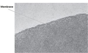
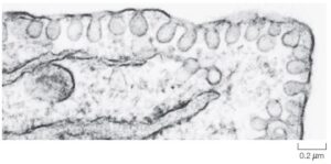
- To investigate plasma membranes more in detail, mammalian red blood cells were used as models for studies, because they lack nuclei and other organelles so they represent a pure source of the plasma membrane.
- The bilayer structure of the erythrocyte plasma membrane, clearly evident in high magnification electron micrographs.
- In lipid bilayer, lipid molecules constitute about 50% of animal cell membrane’s mass excluding protein. In the 1mm x 1mm area of a lipid bilayer, 5 x 10⁶ lipid molecules.
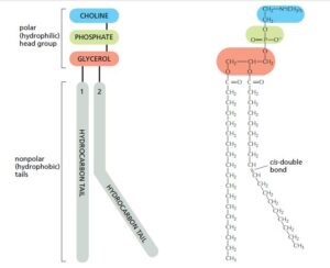
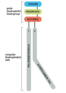
- Lipid molecules are amphiphilic in nature i.e., they have a hydrophilic (“water-loving” or polar end) and a hydrophobic (“water-fearing” or non-polar end).
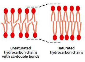
- Phospholipids that are abundant in membrane lipids have a polar head group and two hydrophobic hydrocarbon tails (fatty acid). These hydrophobic tails can differ in length (normally contain 14 – 24 C- atoms), in animals, plants, and bacterial cells.
- One tail is unsaturated (one or more cis bonds), while another tail is saturated. Each cis bond forms kink (bent like form) in the tail. Differences in saturation of fatty acid tails and length influence the packaging of phospholipid molecules, which affects the fluidity of the membrane.
- Major lipids in cell membranes are phosphoglycerides, sphingolipids, and sterols.
- Phosphoglycerides are the main phospholipids of most animal cell membranes, have a three-carbon glycerol backbone. Two long-chain fatty acids are linked by ester bonds to adjacent C- atoms of glycerol and the third C- atom is attached to a phosphate group.
- Different phosphoglycerides are formed by cells by combining different fatty acids and head groups such as phosphatidylserine, phosphatidylcholine, and phosphatidylethanolamine.
- Phospholipids are synthesized in the endoplasmic reticulum and symmetrically arranged themselves in the inner monolayer of membranes.
- Depending on the condition and nature of lipids, it aggregates in three forms,- micelle, bilayer, and liposomes, all structures minimize the contact between a hydrophobic region of lipids with water.

- Another phospholipid that is important is called sphingomyelin, built from sphingosine rather than glycerol backbone like phosphoglycerides. Sphingosine is a long acyl chain with two hydroxyl groups (OH) at one end of the molecule and an amino group (NH2).
- Sphingomyelin is synthesized in the Golgi apparatus, determines its location outer portion of the monolayer
- In sphingomyelin, a fatty acid tail is attached to the NH2 group, and a phosphocholine group is attached to the terminal OH group, remaining free OH group contributes to polar properties of the adjacent head group (as it can form H- bond with the head group of adjacent lipid with a water molecule or with membrane protein).

- In addition to phospholipids, plasma membranes also contain glycolipids and cholesterol.
- Cholesterol is a major constituent of animal cells membrane, it has a rigid ring-like structure, inserted into a bilayer with its OH group close to phospholipid head group, it has a higher affinity for sphingomyelin than other phospholipids, therefore concentrated in the outer leaflet.
- Cholesterol enhances the permeability-barrier properties of the lipid bilayer.


Asymmetry distribution of the Lipid Bilayer
- In two halves of the membrane bilayer, phospholipids are asymmetrically distributed.
- In outer monolayer of the membrane consist mainly of phosphatidylcholine and sphingomyelin, whereas in the inner monolayer, phosphatidylethanolamine and phosphatidylserine are predominant.
- The head group of phosphatidylserine is negatively charged which results in a net negative charge in the cytosolic face of the plasma membrane.
- Transporters are responsible for selectively translocate the phospholipids, and determines their asymmetric orientations.
- In apoptosis, phosphatidylserine translocates to the outer monolayer of the membrane, it signals the elimination of dying cells which is carried out by phagocytosis.

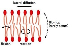
The bilayer is a viscous fluid, long hydrocarbon chains of fatty acids move freely inside the inner plane of the membrane, both proteins and lipids diffuse laterally within the membranes, or rotates on its plane or flip flop which rarely occurs.
- The fluidity of lipid bilayer is essential for normal cell growth and its function depends upon its composition and temperature. An increase in temperature, elevate fluidity of the membrane, whereas low-temperature causes a more rigid, gel-like organization of the membrane, called a phase transition, and here temperature is called transition temperature.

- Bacteria, yeasts, and other organisms adjust fatty acid composition by producing more cis double bonds (it produces kinks, which make them difficult to pack together and form a gel in bilayer), to maintain bilayer fluidity.
- Lipid composition affects fluidity, membranes containing more unsaturated fatty acids are more fluid, the addition of cholesterol decreases the mobility of outer portions of phospholipid fatty acid chains.
Lipid Rafts
- Specialized lipid membrane domains are formed by clustering of a variety of membranes proteins, which are restricted to diffuse freely. Cholesterol, glycolipids, and sphingolipid tend to cluster in small semisolid patches known as lipid rafts.

- These are small transient structures mainly 10–200 nm in size, serves as platforms in which specific proteins can be concentrated and can facilitate their interactions.
- Lipid rafts are enriched in GPI- anchored proteins, and transmembrane proteins involved in a variety of cellular functions which include, cell signaling, cell movement, and endocytosis.
- Interaction of lipid rafts with cytoskeleton results in stabilization of lipid raft and further divide into specialized domains.
- There are two types of lipid rafts- caveolae and planar.
- Caveolae are a subset of lipid rafts, 60- to 80-nm in size and it appears as invaginations of plasma membranes like a flask shape under the electron microscope.
- With the interaction of cytoplasmic protein caveolae and oligomers of membrane protein caveolin, lipid raft caveolae are formed.
- For the formation of caveolae, high content of membrane cholesterol is required, which interacts with membrane protein caveolin.
- Different cell types vary in the number of caveolae, some cells have an abundant number of caveolae, such as endothelial cells and adipocytes, whereas some cells lack caveolae.
- Caveolae have a variety of cellular functions, which include cell signaling, endocytosis, regulation of lipid transport, and protection of plasma membrane against mechanical stress.
- Planar Lipid Rafts- are found in neurons, which contain membrane protein flotillin.
Glycolipid
- Lipid is covalently attached with carbohydrates which can either be monosaccharides or oligosaccharides, it is called glycolipids.
- It can be derived either from sphingosine or glycerol.
- Glycolipids are found exclusively in the outer monolayer of the plasma membrane, with the carbohydrates group exposed on the cell surface.
- E.g., of simplest glycolipid, is cerebroside, contain monosaccharides such as glucose or galactose.
- E.g., of complex glycolipid is globoside, contain oligosaccharide.
Reference and Sources
- https://www.ncbi.nlm.nih.gov/books/NBK9898/
- https://www.flashcardmachine.com/cell-bio-test3.html
- https://www.coursehero.com/file/20928311/Chapter-10-Cell-Memebrane-Structure/
- https://quizlet.com/gb/563807190/internal-organization-of-the-cell-membranestructure-i-and-ii-flash-cards/
- https://quizlet.com/8391717/cell-bio-l4-flash-cards/
- https://oneclass.com/class-notes/ca/york/biol/biol-2021/528056-biol-2021-lecture-2.en.html
- https://courses.lumenlearning.com/boundless-ap/chapter/cell-membranes-and-the-fluid-mosaic-model/
- http://mcb.berkeley.edu/courses/mcb110/Albertsch10.pdf
- https://www.ncbi.nlm.nih.gov/pmc/articles/PMC1304058/
- https://brainly.in/question/8502569
- https://alg.manifoldapp.org/read/fundamentals-of-cell-biology/section/63bdf811-2ef8-41c1-a07bef62d182b7f1
- https://www.cell.com/trends/neurosciences/comments/S0166-2236(02)02215-4
- http://e-portal.ccmb.res.in/e-space/amit/Group%20Publications/2016%20Chol%20200%20review.pdf
This is really very useful and perfect to add few more points for my notes.
Thanks a lot for uploading it.
Thanku, hope it was helpful:)