
E.coli-Epidemiology, Pathogenesis, and Treatment
Introduction
- E.coli belongs to the family Enterobacteriaceae, gram-negative rods, lactose fermenting (produces indole), facultative anaerobes.
- E.coli are ubiquitous in nature, commonly found in water, soil, and as commensal in normal intestinal microflora ( also are opportunistic pathogens).
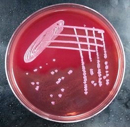
- The culture media used for E.Coli are MacConkey agar, and Blood Agar (Both are Selective Media).
- There are 150 different O antigens & a diverse number of other species (denoted by number for e.g., serotypes antigenic formula denoted by using O, K, and H & antigen present in number such as O111: K76: H7 ).
- Pili is usually present in E.Coli strains, helps to mediate attachment to the epithelial cells of the host.
- Type 1 Pili–—–> interact with the epithelial cell surface ( on D- mannose residues).
- P pili ( known as Pap or Gal-Gal)——>binds to di-galactoside moieties.
- Bundle forming pili (BFP) or Colonisation factor antigens——–>binds to the enterocytes.

- Antigenic structure of E.Coli
- E.coli strains produce four types of toxins that have a cytotoxic effect on the host cells.
-
- α-hemolysin- Pore-forming cytotoxin
- Shiga toxin- AB-type toxin
- Labile toxin (LT)- AB-type toxin
- Stable toxin (ST)- Small Peptides activate membrane-bound guanylate cyclase.
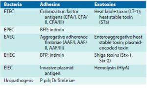
- Virulence Factor associated with E.Coli strains
-
Epidemiology
- A diverse number of E.Coli are present in gastrointestinal microflora and act as an opportunistic pathogen.
- Because of effective virulence factors, it causes various gastrointestinal diseases.
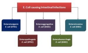
- The five E.Coli pathogenic strains causing intestinal infections or gastroenteritis which lead to diarrhea are ETEC, EAEC, EIEC, EPEC, and EIEC.
- Some extraintestinal infections are also caused by E.Coli strains
- Urinary Tract Infection (UTI)
- Neonatal Meningitis
- Septicemia

Pathogenesis
Enterotoxigenic E. coli (ETEC)
- Commonly observed in children of younger ages.
- Inoculum of the disease is high in number, primarily caused by fecal contamination of the food or water.
- Cause secretory diarrhea, the incubation period is 1-2 days, can persist for an average of 3 to 5 days.
- Symptoms: Watery diarrhea & abdominal cramp, sometimes nausea & vomiting
- The site of infection is the small intestines.
- Two types of Enterotoxin are produced by ETEC:
- Heat-labile Toxins (LT-I, LT-II)
- Heat Stable Toxins (STa and STb)
- LT-II is not related to human diseases and LT-I is an AB toxin, which consists of 1 subunit of A and 5 subunits of B
- In LT-I toxin, B subunits bind to receptors as GM1 gangliosides and other glycoproteins present on epithelial cells.
- While A subunit moves to cross the vacuole membranes, binds with Gs proteins that control adenylate cyclase.
- Because of interaction, cyclic adenosine monophosphate (cAMP) levels increase, which causes more secretion of chloride, and absorption of sodium and chloride is decreased.
- These modified alterations lead to watery diarrhea and also triggers the secretion of prostaglandin & produce inflammatory cytokines, which results in further water loss
- On the other hand, heat-stable toxin STa is a small peptide that interacts with the transmembrane guanylate cyclase receptors, which leads to the secretion of cGMP ( cyclic guanosine monophosphate ) which cause fluid hypersecretion.
Enteropathogenic E.Coli (EPEC)
- Cause infant diarrhea is commonly observed in poor countries.
- Infectious disease is low, person-to-person transmission occurs.
- Symptoms: Watery diarrhea, fever, and vomiting may be present.
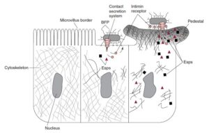
- Initially, bacteria get attached to small intestine epithelial surface cells along with the disruption of the microvillus (known as effacement), which gives a lesion on the microvillus known as Attachment/ Effacement [A/E] histopathology.
- First, microcolonies on the epithelium surface cells are formed by plasmid-encoded bundle forming pili (BFP).
- Subsequent attachment is carried out by genes present pathogenicity island known as “locus of enterocyte effacement”.
- Consists of more than 40 genes that carry out the attachment & destruction of the host cell.
-
- Bacterial type III secretion system, helps bacteria to secrete active proteins in the host cells.
- One protein, Tir (translocated intimin receptors) is inserted on the epithelial cell for bacterial adhesin.
- The binding of Tir receptors and intimin results in actin polymerization and cytoskeletal elements accumulation, under the attached bacteria, which cause cell death.

Enteroaggregative E. coli (EAEC)
- Cause persistent watery diarrhea along with dehydration in infants.
- Cases reported in the US, Europe, Japan, and other developing countries
- Some E.Coli strains can cause chronic diarrhea which also causes retardation in the infant.
- The characterization of the bacteria is done by autoagglutination.
- Its mediated by AAF1 (The bacteria are characterized by their autoagglutination adherence fimbriae I, adhesins).
- EAEC attaches to the intestine surface, which stimulates mucus secretion and leads to thick biofilm formation.
- Biofilm protects aggregated bacteria from antibiotics & phagocytic cells.
- EAEC produces enteroaggregative heat-stable toxic and plasmid-encoded toxin which induces secretion of the fluid.
Enterohemorrhagic E. coli (EHEC)
- EHEC is the common E.Coli strain, causes gastrointestinal disease in developed countries.
- The infection reported is 73000 cases/ yr, mortality reported per year is 60 cases in the US.
- Caused in warm months, affecting children under 5 yrs.
- Usually, consumption of undercooked beef, or other meat products, unpasteurized milk, uncooked vegetables can lead to the cause of infections.
- Inoculum dose is low, even 100 bacteria can cause disease, transmission can take place by person-to-person contact.
- Symptoms: Mild diarrhea to hemorrhagic colitis which can cause bloody diarrhea ( known as dysentery) and sharp abdominal pain, in a few patients vomiting can be observed.
- The incubation time period is 3 to 4 days, symptoms like diarrhea & abdominal pain are developed.
- 30% to 65% of patients experience bloody diarrhea & severe abdominal pain at the onset of 2 days.
- EHEC is related to HUS (Hemolytic uremic syndrome), which leads to acute renal failure, deficiency of the platelets ( thrombocytopenia), intravascular hemolysis ( microangiopathic hemolytic anemia), these symptoms complicate the case.
- EHEC serotype, such as O157:H7 is a common strain to cause disease
- EHEC produces Shiga toxins (Stx-1 and Stx-2, ), both are AB-type toxins,
- Here B subunits bind to the specific glycolipid such as globotriasylceramide[Gb3] present on the host renal endothelial cell and intestinal villi.
- A subunit is internalized, and cleave into two molecules, one part binds to 28S rRNA and ceases protein synthesis.
- EHEC strains are more pathogenic if it has both Shiga like a toxin and Attaching and Effacing activity.
Enteroinvasive E. coli (EIEC)
- The occurrence of disease caused by EIEC is rare both in developing & developed countries.
- Few serotypes of EIEC strains are pathogenic such as O124, O143, and O164.
- Bacteria invade and cause destruction in the colonic epithelium. (Associated with large intestines).
- Symptoms: Watery diarrhea, may progress into dysentery, fever, abdominal cramps, and blood and leukocyte in the stool samples.
- Bacterial invasion is mediated by pInv genes.
- Bacteria cause lysis of phagocytic vacuoles and multiply in the cytoplasm.
- The formation of actin tails, helps bacteria to move within the cytoplasm and across the neighboring epithelial cells.
- Colonic ulceration occurs because of epithelial cell destruction with inflammatory cytokines stimulated by bacteria
| Escherichia coli (Strains) | Site of Infection | Disease | Remarks |
| Enteropathogenic E. coli (EPEC) |
Small Intestine | Classic infant diarrhea | Epidemics in hospitals, children’s homes |
| Enterotoxigenic E. coli (ETEC) |
Small Intestine | Traveler’s Diarrhea | Cause of travelers’ diarrhea ( 50 %) |
| Enteroaggregative E. coli (EAEC) |
Small Intestine | Watery diarrhea, mainly in infants |
Adhesion to the small intestine mucosa; production of a toxin |
| Enterohemorrhagic E. coli (EHEC) |
Large Intestine | Hemorrhagic colitis | Hemolytic–uremic syndrome (HUS) in 5% of EHEC cases |
| Enteroinvasive E. coli (EIEC) |
Large Intestine | Dysentery | Invasion and verocytotoxins |
Diagnosis
- Immunoassay and nucleic acid detection can be used to determine the toxins and their associated genes. It’s expensive to be used practically for diagnosis.
- MacConkey agar is used as a culture medium( Pink colonies is obtained), for EHEC in media instead of lactose, sorbitol is incorporated. (Colourless Colonies is obtained)
Treatment
- Rehydration and supportive measures are taken into consideration.
- Hemorrhagic colitis and HUS are treated by using hemodialysis or hemapheresis.
- To treat diarrhea caused by ETEC, EIEC, and EPEC, trimethoprim/sulfamethoxazole (TMP-SMX), doxycycline, or quinolones.
References and Sources
- https://quizlet.com/292896388/unit-5-gi-flash-cards/
- https://www.researchgate.net/publication/5850838_Escherichia_coli_STb_toxin_and_colibacillosis_Knowing_is_
half_the_battle - https://www.sciencedirect.com/topics/nursing-and-health-professions/methyl-red
- https://quizlet.com/289127010/micro-final-diagram/