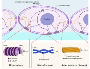
Cytoskeleton Definition
- Cytoskeleton is Intracellular network of protein filament present in cytoplasm.
- It is composed of three different types of proteins which are organized and regulated in time and space.
- It is easily recognizable.
- These different types of cytoskeletal fibers are polymers which are made up of small protein subunits and held together by non-covalent interactions.

Cytoskeleton Functions
- It decide proper positioning of organelles.
- It provide track for vesical transport which helps in exocytosis and endocytosis, and also play important role in intracellular vesicular transport.
- It provide proper shape of the cell.
- It is helpful cellular movement.
- It also involve in contractibility and motility.
| Feature | Microtubule | Intermediate filament | Microfilament |
| Diameter | Approx 25 nm | 10-12 nm | 7-8 nm |
| Monomer | Alpha and Beta tubulin | More than 70 different protein | Actin ATP |
| Polarity | Present | Absent | Present |
| Motor protein | Kinesin and Dynein | No motor | Myosin |
| Strength | Tensile and Hollow | Filamentous and Rigid | Solid and rigid |
| Regulation of assembly and disassembly | Assembly and disassembly regulated by phosphorylation and dephosphorylation | By phosphorylation and dephosphorylation | Regulated assembly by large number of protein and disassembly |
| Dynamic | Highly | Less | Highly |
| Functions | Provide organisational framework for organelle transport, it support cilia and flagella and form mitotic spindle. | It support nuclear envelope (lamin), cells and tissue integrity, support exoskeleton. | It support plasma membrane and involve in formation of microvilli and pseudopodia, acts as contractile machinery and network for the cell cortex. |
Types of Cytoskeleton
Microtubule
- It is Composed of 13 proto filament which are laterally associated with each other and form hollow core.
- Each proto filament is made up of Alpha and Beta tubulin and both subunits are covalently linked.
- Alpha and beta tubulin are heterodimer and arranged in head to tail manner, which provide polarity to microtubule that means, at positive end beta tubulin is exposed and at negative end alpha subunits is exposed.
- The positive end is fast growing, while negative end is slow growing end.
- Each alpha and beta tubulin bind with GTP, but GTP found in beta tubulin can be hydrolyse during polymerization.
- Microtubule assembly is catalysed by optimum temperature at 37o.
- Assembly required MAPs (microtubule associated proteins) which stabilize the microtubule.
- Tubule polymerization involved three steps:
- Nucleation phase
- Elongation phase
- Steady state
Stabilization of microtubule
- Tau family of proteins MAP-2 and MAP-4 having positively charged amino acids which binds to negative charged tubulin surface and stabilize the microtubule.
- Drugs- colchicin, colcimid, podophylotoxin, vincristine block the microtubule polymerization.
- Taxal inhibit microtubule depolymerisation.
- Two family of motor protein associated with microtubule:
- Kinesin
- Dynein
- Two family of motor protein associated with microtubule:
- Kinesin
- Kinesin is plus end directing motor protein and it is tetrameric in nature.
- Kinesin-1 is responsible for anterograde pathway.
- Kinesin-14 , it is only kinesin which moves at negative end of microtubule.
- Kinesin-13 , it is microtubule depolymerase, no motor activity, it binds with positive end of tubule and trigger depolymerisation, play important role in metaphase to anaphase transition.
- Dynein
- Minus end directing motor protein.
- It is engaged in retrograde pathway.
- Dynein link cargo by using dynectin protein.
- It play important role in chromosome movement during mitosis.
Intermediate filament
- It is found only in animals, the Intermediate filaments are more stable and interconnected to cytoskeletal element by the cross bridges and using plectin.
- No any ATP and GTP required for assembly and disassembly.
- Assembly and disassembly regulated by phosphorylation and dephosphorylation e.g. Lamin protein.
- Keratin of epithelial cells, desmin and vimnectin of muscle cells, fibroblasts and WBC, neurofilament of neurons, lamin of nucleus are main intermediate filament.
- Keratin is more diverse protein and categorized into two classes.
- Hard keratin- having high disulphide bond and rigid in nature, E.g., hair and nail.
- Desmin protein- It connects sarcomere and play important role in contraction.
- Lamin- It is component of nuclear lamina.
- Soft- keratin/cytokeratin- it is associated with desmosomes and makes cell to cell contact.
Microfilament (actin filament)
- G- actin ATP is monomer of microfilament .
- Actin polymerization is also completed into 3 steps:
- Nucleation phase
- Elongation phase
- Steady state
- Actin filament also show polarity that means, it represents positive end and negative end.
- Actin filament trade miling is regulated by two class of proteins, profilin and cofilin.
- Profilin trigger polymerization and cofilin trigger depolymerisation.
- The remodelling of actin cytoskeleton is required for cell membrane for cell movement, the changes in the cell shape, phagocytosis, cytokinesis etc.
- Capping protein block the assembly and disassembly of actin filament
- Cap-z protein having high affinity for positive end of actin filament, that’s why after binding of Cap-z to the positive end ,prevent addition or loss of actin at positive end.
- Tropomodulin having high affinity for negative end, that’s why it inhibits assembly and disassembly of negative end.
- Cytochalacin-D and latrunculin promote actin depolymerisation.
Myosin
- It is main motor protein of actin filament.
- 14 myosin families are identified that regulate muscle physiology by using actin protein.
- Myosin is of two types:
- Conventional myosin: This type of myosin present in muscle cells. E.g. Myosin 2.
- Non-conventional: Present in non-muscle cells, e.g. myosin 1 – regulate cargo vesicle move towards the plus end. Myosin 3- it having sensory role in vision. Myosin 6 and 7 play important role in maintenance of sensory mechanism in hearing.
- The contractile assembly of actin and myosin occurs in muscle as well as in non-muscle cells.
- It regulates formation of contractile ring during cytokinesis.
Reference and Sources:
- https://www.mechanobio.info/cytoskeleton-dynamics/what-is-the-cytoskeleton/what-are-actin-filaments/whatis-actin-nucleation/
- https://bio.libretexts.org/Courses/University_of_California_Davis/BIS_2A:_Introductory_Biology_(Easlon)/Readings/14:_The_Cytoskeleton
- https://www.khanacademy.org/science/biology/structure-of-a-cell/tour-of-organelles/a/the-cytoskeleton
- https://onlinelibrary.wiley.com/doi/abs/10.1002/cm.970300203
Also Read:
- Gram staining
- DNA Replication in eukaryotes: Initiation, Elongation and Termination
- Shigella-Epidemiology, Pathogenesis, and Treatment
- Tetanus – Etiology, Pathogenesis, and Treatment
- The Nucleus-Detailed Structure and Functions
- Beta Variant: Introduction, Symptoms, Vaccines and Prevention
- Enzymes: Introduction, Enzyme activity and work Mechanism
- Cell Cycle: Introduction, Phase, Mechanism and Significance

