Introduction
- In multicellular organisms, growth, development, and maintenance depends not only on the production of cells but it also depends on the mechanism to destroy them.
- For the maintenance of tissue size, it requires that cells die at the same rate as they are produced.
- Death of cells also occurs when the cells are infected or damaged, ensuring that they are eliminated before they threaten the health of organism.
- Death of cell is not random process, it occurs by a programmed cascading molecular events, in which cell destroys itself systematically from within and is engulfed by other cells, leaving no trace.
- In most of the cases, this systematic programmed cell death occurs by a process termed as apoptosis.
- Apoptotic cells are engulfed or eliminated by phagocytic cells such as macrophages.
- All cells having limited lifespan and completing their life span very cell enter into apoptotic phase.
- Apoptosis is a natural phenomenon which leads natural death of cell.
- If abnormality found in apoptosis then the lifespan of cell is increased and promotes cancer development.
- All multicellular organisms require external factors that provide signal to stay alive and those factors are called tropic factors.
- In absence of trophic factor or survival signal cell activates suicidal program.
- Apoptosis involves elimination of cells that having irrepairable DNA damage.

Two form of cell death
Brief distinguish features of necrosis and apoptosis
| Necrosis | Apoptosis |
|
It is programmed cell death, death by suicide. |
| Energy dependent. | |
|
In this unwanted cells are eliminated by systematic biological process. |
| Phagocytosis involved. | |
| Apoptosis may be through intrinsic or extrinsic pathways. | |
| Apoptotic events involves, cell becomes more compact, blebbing of membrane, chromatin condensation, DNA fragmentation, and cell shrink, and release of apoptotic body. | |
|
Regulators of Apoptosis
- Bcl2 a mammalian gene which was first identified as oncogene that involved in the development of cancer of B- Lymphocytes.
- It was found to inhibit apoptosis.
- Bcl2 family of proteins are characterized by the presence of one or more small BH domains.
- There are approximately 20 proteins related to bcl2 which was encoded by mammals and these are divided into three functional groups including:
- Anti-apoptotic: it protects the cells from apoptosis, gain of function mutation in these genes, increases cellular lifespan thus causes cancer. E.g. Bcl-xl, Bcl-w and Bcl-2.
- Pro-apoptotic: it promotes apoptosis, loss function mutation in these genes, inhibits apoptosis, increases cell life span and causes cancer. E.g. Bax and Bak.
- BH only: it promotes apoptosis by indirect mechanism. E.g. Bid, Bad, Puma.
Examples of anti-apoptotic and pro-apoptotic factors
| Anti-apoptotic factors | Pro-apoptotic factors |
|
|
Caspases
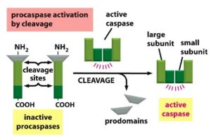
- Caspases are group of proteases which have cysteine residue in their catalytic site and cleaves protein at the C- terminal of aspartate residues.
- Caspases are cysteine dependent aspartate specific protease.
- Caspases are ultimate effector of programmed cell death and involved in the apoptosis by cleaving more than 100 different target proteins.
- Caspases are inhibitors of DNase, which on activation responsible for the fragmentation of nuclear DNA.
- It also involves in the cleavage and activation of scramblase.
- In addition caspases cleaves cytoskeletal proteins which results in the cytoskeleton disruption, irregular bulging or blabbing of plasma membrane and cell fragmentation.
- Involves in the fragmentation of nucleus by cleaving nuclear Lamin protein.
Types of caspases
Initiator caspases:
-
- It activated by self autocatalysis.
- Begins the process of apoptosis.
- E.g. caspase- 2,8,9,10.
Effector caspases:
-
- It get activated by the Initiator caspases.
- E.g. caspase- 3,6,7.
- All caspases are synthesized in an inactive form known as procaspases, which are then converted into active from by cleavage of proteins catalysed by other caspases.
- Caspases deficiency may results in the tumour development.
- In animals, caspases are responsible for the apoptosis, while in plants and fungi, apoptosis induced bg arginine and lysine specific caspase such as metacaspase.
- Not all caspases activates apoptosis.

Apoptotic pathways
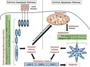
Intrinsic pathway/ cell suicide program
- This pathway is favoured in the absence of trophic factor.
- Activation of intrinsic pathway is regulated by membrane bound bcl2 family protein which anti-apoptotic in nature and behave as a oncogene by promoting survival of the cell.
- In absence of trophic factor PK-B (protein kinase-B) is inactive that’s why Pro-apoptotic bad protein is active.
- Active Bad protein bind with Bcl-2 protein and cause its activation.
- If Bcl-2 is inactive then, Pro-apoptotic Bax is polymerised and behave as a channel, through which cyt-c exported from IMS site to cytosol.
- In cytosol it binds with Apaf-1 and form wheel like, hepatmeric structure called apoptosomes.
- Apoptosomes activates caspase 9 which is initiator caspase.
Extrinsic pathway/ murder program
- It occurs through the death signal.
- TNF-alpha released by macrophages and triggered cell death.
- Fas-ligand is a cell surface protein produced by activated natural killer cell and Tc cell.
- These signals trigger cell death of infected cells and foreign grafted cells.
- Murder signal comes from outsides of the cells and binds to cell surface receptor termed death receptor.
- Death receptor is trans-membrane protein containing extracellular ligand binding domain and intracellular death domain which is required for receptor to activate apoptotic programme.
- In cancerous cells death receptors are mutated.

Biochemical changes during apoptosis
Cell undergoes apoptosis having certain biochemical changes, which can be used as indicator or marker of the apoptotic cell.
- Negatively charged phosphatidyl serine located at cytosolic leaflet of the membrane, but during stressful condition it flip out to exoplasmic leaflet in ATP dependent manner where it recognize as eat-me-signal or marker of the death signal.
- Anexin-5 located at extracellular domain and it specifically binds to phosphatidyl serine and activates professional phagocytic cells like macrophages and dendritic cells.
- Nuclei is condensed, DNA is fragmented by topoisomerase and protein is cleaved by protease, cleavage of DNA marked as late apoptotic nuclei or necrotic nuclei.
- Nuclear condensation promotes nuclear shrinkage, cell shrinkage and forms small membrane bound apoptotic body, which is engulfed by phagocytic cells.

Reference and Sources:
- https://en.wikipedia.org/wiki/Apoptosis
- https://www.ncbi.nlm.nih.gov/books/NBK26873/
- https://quizlet.com/156316283/cell-death-and-apoptosis-flash-cards/
- https://www.khanacademy.org/science/biology/developmental-biology/apoptosis-in-development/a/apoptosis
- ttps://www.sciencedirect.com/topics/neuroscience/protein-bcl-2
- https://www.nature.com/articles/s41419-020-2589-7
- https://quizlet.com/172840400/chapter-18-apoptosis-programmed-cell-death-flash-cards/

