
Bacteria: Shape, Size, Structure, and other Membrane
Introduction
- Members of domain bacteria are microscopic, relatively simple, prokaryotic organism that lacks a nucleus.
- Bacteria are usually single celled organisms.
- Peptidoglycan is present in eubacterial cell walls.
- Bacteria have an ability to grow on artificial laboratory media.
- Reproduction is asexual type.
- Some Bacteria have ability to cause diseases, some form important role in natural cycling of elements which contribute to soil fertility.
- Bacteria also useful in industry for manufacture of valuable compounds, manufacturing of antibiotic, food products, some spoil foods and some make foods.
Size of Bacteria
- Bacteria range in size from 0.2 – 2 micrometers in diameter, and 0.5 – 5.0 micrometers in length.
- Thiomargarita namibiensis & Epulopiscium fishelsoni are large in size and visible through the naked eye.
- mycoplasma is the smallest bacteria.
Shape and arrangements of bacterial cell
Cell shape is generally characteristics of a given Bacteria species. The four basics shapes of Bacteria are:
- Rod like – bacillus.
- Spherical or ovoid- coccus.
- Comma shaped- vibrio.
- Spiral or helical shaped- spirilla.
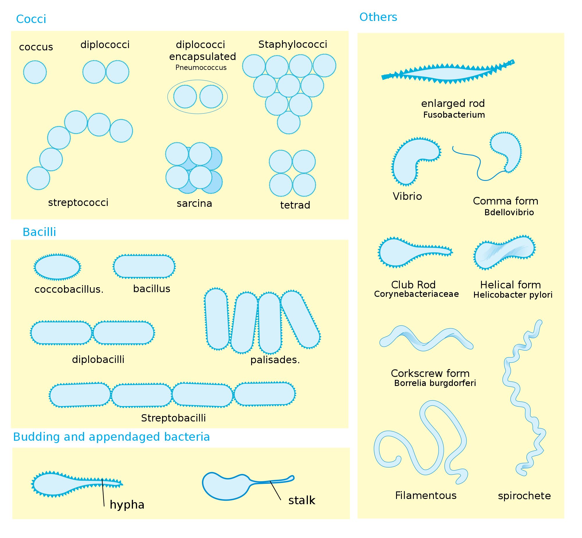
- When cocci divide, cell can remain attached to one another.
- Diplococci remains in pairs, cells divide in one plane.
- Streptococci remain attached to form chains and cells divides in one planes.
- Tetrad cocci divides in two planes and remains in group of four, forming square.
- Sarcinae are termed when it divides in three planes and form cubelike structure.
- Those that divides and form grape like clusters are called staphylococci.
- Diplobacilli appears in pairs after division and streptobacilli occurs in chains.
- Spirilla fairly rigid Bodies and helical shape.
- Spirochetes spirals which are helical and flexible.
- Most Bacteria maintain single shape are termed as monomorphic.
- Some bacteria like corynebacterium, many shapes and lacks a single, characteristics shape termed as pleomorphic.
Structure of Bacteria
Cell wall
- Bacterial cells almost always bounded by a chemically complex wall.
- The main function of cell wall is to protect bacteria from osmotic lysis.
- Cell wall is chemically composed of peptidoglycan (also termed as Murein).
- In gram positive Bacteria, peptidoglycan contains 20 to 80 nm thick homogeneous layer present outer side of the plasma membrane.
- The gram negative bacteria consists of 2 to 7 nm thick peptidoglycan layer with a 7 to 8 nm thick outer membrane.
- Peptidoglycan is a polymer which contains two sugar derivative:
- N-acetylglucosamine (NAG).
- N-acetylmuramic acid (NAM).
- Amino acids present in tetra peptide includes L- alanine, D- alanine, D- glutamic acid, and either L- lysine or mesodiamino-pimelic acid (DAP).
- Many gram negative bacteria have acidic substance called teichoic acids in their cell wall.
- The important function of teichoic acid is to give rigidity to the cell wall by attaching cation like magnesium and sodium.
Outer membrane
- In addition to peptidoglycan cell wall, the gram negative bacterium contains an additional membrane, the outer membrane.
- It contains lipopolysaccharide, lipoprotein, protein and phospholipids.
Glycocalyx
- Glycocalyx is thick, high molecular – weight secretory substance and is present in many Bacteria external to the cell wall.
- The rigid layers are organized in a tight matrix called a Capsule.
Surface appendages
Three types of surface appendages:
- Flagella for locomotion.
- Pilli for conjugation.
- Fimbriae for attachment.
Flagella
- Bacterial flagella are hairlike, helical appendages that protrude through the cell wall and responsible for motility or locomotions.
- Flagella occurs in both grams positive and gram negative bacteria.
- A flagellum of a gram negative Bacteria such as E. coli, contains three parts:
- The long helical filament, which is present on the cell surface.
- The short hook structure is present at the end of the filament.
- The basal body, to which the hook is embeded and which helps in motion to the flagellum.
Number and distribution of flagella:
- Monotrichous have one flagellum.
- Amphitrichous one flagellum attached at end.
- Lophotrichous cluster of flagella at one end or both ends.
- Petritrichous flagella are spread fairly on whole surfaces or surrounded by lateral flagella.

Pilli
- The term pilli is used to describe the thin, hairlike appendages on the surface of Bacteria.
- Pilli is generally present in gram negative Bacteria.
- pillins is the protein that form pilli.
- It plays important role in the process of conjugation.
- It also function as a receptor for donor specific phages.
Fimbriae
- Fimbriae are filamentous cell appendages.
- It is present in gram positive as well as in gram negative bacteria.
- Mainly arranged by helically composed protein subunit.
- Fimbriae mediate cell attachment to the surface.
Reference and Sources
- 1% – https://courses.lumenlearning.com/wmopen-biology2/chapter/the-structure-of-prokaryotes/
- 1% – https://www.sciencedirect.com/science/article/pii/B9780128174951000074
- 1% – https://www.biologydiscussion.com/notes/bacteria-notes/notes-on-bacteria-biology/34260
- 1% – https://alldokument.com/micro-biology-std11-english-medium.html
- 1% – https://quizlet.com/224428733/microbiology-chapter-4-flash-cards/
- 1% – https://www.ncbi.nlm.nih.gov/pmc/articles/PMC3359400/
- 2% – https://www.slideshare.net/sardar1109/bacteria-64730553
- 1% – https://pt.scribd.com/document/155465203/5097
- 1% – https://www.studyblue.com/notes/note/n/exam-2/deck/4346216
- 1% – https://idoc.pub/documents/thephysiologyandbiochemistryofprokaryotes-1430rzprgo4j
- 1% – https://www.ncbi.nlm.nih.gov/books/NBK8477/
- 1% – https://www.cell.com/trends/microbiology/comments/S0966-842X(99)01513-9
Also Read:
- Proteins: Definition, Roles, Functions and Structure
- Bacterial vaccines – An Overview
- Gram staining
- Mycobacterium tuberculosis-Epidemiology, Pathogenesis, and Treatment
- Difference between Prokaryotes and Eukaryotes